


A session ofiridology
What to Expect During a session ofiridology
Historical Foundations and the Iris Study Model
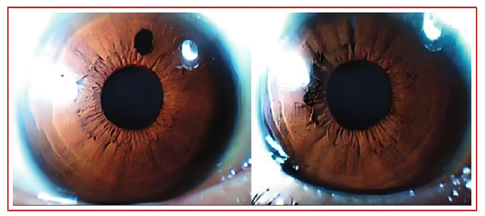
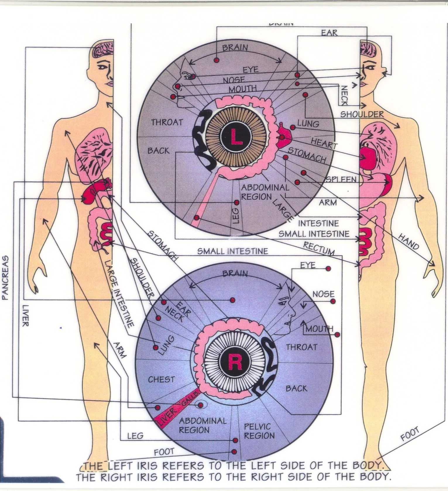
Iridology developed from the premise that micro-structural variations within the iris correspond to various parts of the human anatomy. The 19th-century observations of Hungarian physician Ignatz von Peczely established the earliest topographical grids, suggesting that specific quadrants of the iris reflect systemic stress in different organs systems. Modern holistic health practitioners view the iris as an individualized map that reveals inherited tissue resilience rather than active infections or specific internal injuries.
It is essential to distinguish between regular eye exams performed by optometrists and an iridology study iris assessment. Optometrists inspect internal structures such as the retina, optic nerve, and crystalline lens to identify vision impairments and ocular pathologies. In contrast, iridologists observe the colored part eye—specifically the anterior surface of the iris, the pupillary border, and the collarette—solely for holistic constitution profiling.
Observation of the anterior iris stroma provides insight into constitutional tendencies and tissue tone. These findings serve educational purposes and do not confirm, quantify, or rule out medical conditions.
The practice recognises that the pigment distribution, lacunae, and crypts on the surface of the eye remain largely stable throughout adulthood. Rather than signalling immediate disease, these permanent structural characteristics provide clues regarding hereditary stamina and metabolic tendencies. Holistic consultants use these markers to guide long-term wellbeing routines rather than acute medical interventions.
Structuring the Initial Consultation
An effective iridology evaluation begins well before any optical hardware is turned on. A comprehensive initial consultation establishes personal lifestyle context, dietary preferences, environmental stressors, and familial health patterns. Understanding these elements enables complementary therapy professionals and naturopathy practitioners to evaluate iris features within an individual’s real-world environment.
- Detailed personal lifestyle and dietary assessment to identify systemic vitality and daily habits.
- Review of family health history to understand inherited genetic strengths and susceptibilities.
- Discussion of current wellbeing targets, energy patterns, and digestive tendencies.
- Explanation of equipment safety, illumination levels, and photographic procedures.
- Clarification of observational boundaries, establishing that the assessment does not diagnose medical diseases.
Clients are asked to remove contact lenses prior to optical capture, as tinted or reflective corrective lenses can distort the true colour balance and obscure minute trabecular fibres. Eyeglasses are also set aside so that the lens of the optical device can approach the orbital area without hindrance. This preparatory dialogue helps the client remain relaxed, keeping the eyelids steady during digital image acquisition.
Digital Imaging and Iriscope Hardware
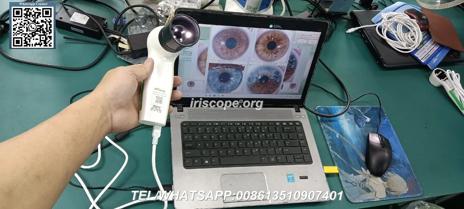
Modern practitioners rely on specialised digital instruments to obtain stable, high-magnification photographs of both eyes. The MAIKONG Iris 12MP High Pixel Professional USB Iriscope Camera (Model GY-9822U) illustrates the optical engineering required to capture clear, distortion-free ocular imagery. Built with an imported coated lens, a dedicated DSP image processor, and optical image stabilization, the instrument delivers 12.0-megapixel digital captures that reveal intricate stromal architecture.
| Specification | Technical Parameter | Operational Significance |
| Sensor Type | 12.0 Megapixel CCD | Captures high-resolution detail of iris fibres and pigment margins. |
| Illumination | Integrated Circular LED | Supplies balanced lighting across the ocular surface without harsh glare. |
| Optical Lens | 30X Iris Lens | Provides optimal focal magnification for mapping structural lacunae. |
| Power Source | Direct USB Connection | Draws power straight from the host computer without external mains adapters. |
| Classification | Class I Instrument | Operates as a safe, low-voltage biochemical and optical analysis tool. |
Circular LED lights positioned around the optical barrel illuminate the iris evenly, minimising the dark shadows cast by the brow ridge or eyelashes. Adjustable focus controls on the handset allow the operator to fine-tune focal depth for differing corneal curvatures, while automatic white balance maintains natural colour rendering. A built-in single capture button with digital pause enables instant image stabilization without shaking the camera body.
Step-by-Step Procedure in a session ofiridology
A systematic operational protocol maintains procedural consistency, client comfort, and accurate photographic documentation. When embarking on a session ofiridology, both practitioner and client benefit from a structured, calm routine that prevents visual fatigue from ocular illumination.
- Host System Setup: The operator connects the USB iriscope to a compatible workstation and verifies software communication.
- Client Positioning: The client sits comfortably with their head balanced, looking straight ahead into the optical barrel.
- Illumination Adjustment: The practitioner switches on the soft LED ring light and adjusts light intensity to prevent squinting.
- Focal Alignment: Using the manual focus wheel on the handset, the operator sharpens the view of the pupillary margin.
- Image Capture: The practitioner presses the capture button to freeze the image, capturing both the right (OD) and left (OS) eyes.
- Data Archiving: Photographs are assigned immediately to the client digital file, indexed with date, time, and session notes.
- Comparative Review: The captured files are projected onto the workstation screen for collaborative discussion and chart overlay.
Each eye is examined separately because the anatomical representation differs between the hemispheres. The right iris corresponds primarily to the right side of the body, while the left iris reflects the left side. Practitioners take multiple exposures if involuntary blinks or reflections obscure key zones, choosing the sharpest image for subsequent evaluation.
Software Mapping and the Iridology Chart
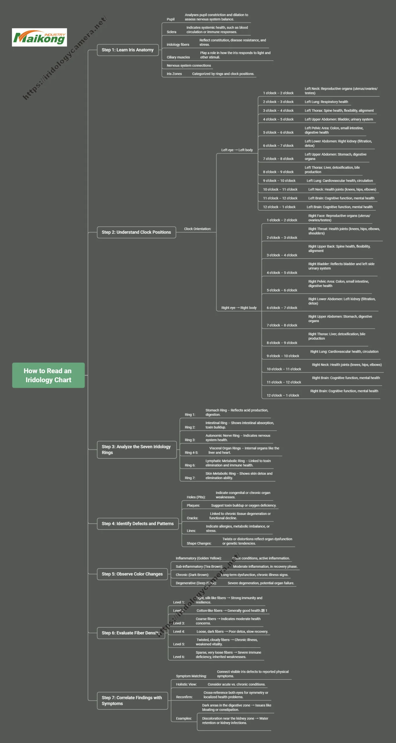
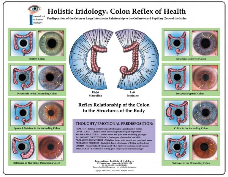
Once high-resolution images are secured, analysis proceeds by superimposing an iridology chart over the digital photograph. The chart divides the annular area of the iris into concentric rings and radial sectors resembling a clock face. The inner pupillary zone typically relates to gastrointestinal processes, the intermediate ciliary zone connects to circulatory and glandular function, and the outer rim corresponds to skin and lymphatic flow.
Digital software overlays allow practitioners to align radial sector grids precisely over variable pupil sizes, assisting in the consistent documentation of individual iris zones.
Modern analytical software simplifies this mapping process by adjusting the digital grid to match varying pupil diameters and decentrations. Programs like MAIKONG 2026 Iridology Software provide 3D texture visualisation, negative-film colour inversion, and multi-date image comparison. These features enable practitioners to demonstrate subtle surface shifts, pigment variations, and constitutional markings directly to the client on screen.
The software also maintains organised client registries, generates visual wellness summaries, and logs personalised lifestyle suggestions. Practitioners can archive successive visits across months or years to assess whether supportive nutritional regimens coincide with visible improvements in tissue vitality.
Interpreting Iris Findings: Health Guidance versus Medical Diagnosis
A central ethical duty in every session is maintaining a clear distinction between holistic evaluation and medical diagnosis. An iridology assessment does not identify specific pathologies, bacterial infections, or named diseases. Signs that practitioners describe as inflammation, toxicity, or lymphatic sluggishness represent qualitative constitutional patterns rather than clinical conditions like high blood pressure, ulcers, or malignancy.
Iridology findings are solely for personal wellbeing education and constitutional evaluation. They do not substitute for professional medical consultations, clinical examinations, laboratory testing, or pharmacological therapy.
When an iridologist notes a concentrated pigment spot, an open lacuna, or radial furrows in a specific sector, they interpret these as areas that may benefit from supportive lifestyle care. The observation does not mean the corresponding physical organ is damaged or failing. Responsible consultants explicitly advise clients to seek formal medical review whenever acute symptoms, pain, or suspicious health changes occur.
By framing findings around vitality, balance, and general organ workload, iridology operates as an empowering educational model. Clients gain appreciation for their baseline physical constitution, encouraging proactive hydration, wholesome nutrition, stress moderation, and restorative rest.
Functional Wellness, Longevity, and Clinic Integration
Modern integrative clinics, spa operators, and longevity centres frequently incorporate iris analysis into broader preventative wellness programmes. When paired with complementary modalities, iris photography provides an accessible visual starting point for dialogue on lifestyle longevity, anti-aging protocols, and cellular vitality.
Nutritional Therapy
Guides dietary adjustments and mineral replenishment based on observed digestive and elimination constitutional indicators.
Detox Protocols
Assists holistic programmes, including sauna or gentle detox regimes, by evaluating lymphatic and cutaneous markings.
Vitality Support
Integrates alongside therapies like ozone or relaxation techniques in a wellness clinic to monitor self-reported rejuvenation.
In multi-disciplinary facilities, iridologists collaborate with herbalists, reflexologists, and health coaches to form holistic care strategies. The iris image serves as a visual touchstone, helping clients follow the reasoning behind dietary recommendations, botanical infusions, or detoxification protocols. The focus remains consistently on promoting constitutional balance, optimising organ reserve, and supporting long-term health.
Technical Specifications and Customisation Systems
Practitioners and wellness centres require reliable hardware and adaptable software tools to conduct professional assessments. MAIKONG manufactures hardware and software systems with extensive customisation options, allowing institutions, academies, and private clinics to deploy consistent diagnostic aids. Professional buyers can source equipment through specialized OEM channels to tailor hardware housings, branding, and packaging to local practice standards.
| System Platform | Supported Architecture | Software Capabilities |
| Windows OS | Win 2000, XP, Vista, 7, 8, 10, 11 (32/64-bit) | Full automatic grid matching, 3D texture, client database, report exports. |
| Apple macOS | macOS 10.14 through macOS 15 (Sequoia) | Integrated ocular analysis, client archiving, high-resolution visual capture. |
| Non-Supported | Linux, Unix, Android | These operating systems are not supported by the software suite. |
The MAIKONG software ecosystem supports 17 distinct language environments, including English, Spanish, French, German, Italian, Portuguese, and Simplified Chinese. Hardware engineering complies with Class I equipment standards and CE requirements, following ISO9000 quality control processes throughout manufacturing. Custom options include exterior colour schemes, bespoke mould design, and custom software report templates that integrate recommended nutritional ranges.
UK Professional Practice and Data Governance
Conducting iris photography sessions within the United Kingdom involves adherence to established complementary therapy professional standards and data protection principles. Practitioners must safeguard client privacy, obtain informed consent before imaging, and store digital records securely. Because iris images represent unique biometric identifiers, handling digital files with strict confidentiality is essential.
Professional Practice Strengths
- Completely non-invasive procedure offering instantaneous visual client engagement.
- Permanent digital image archiving allows objective comparison over successive appointments.
- Encourages clients to take an active, informed role in personal lifestyle choices.
Operational Considerations
- Hardware requires precise lens positioning and pupil illumination calibration.
- Observations are strictly constitutional and cannot replace validated laboratory diagnostics.
- UK practitioners must establish compliant client data storage and consent records.
When selecting professional devices, clinic owners should review equipment durability, warranty provisions, and technical support access. For information regarding corporate background and engineering quality, practitioners can review the About us documentation from MAIKONG. The company states a typical design lifespan of 5 to 10 years for its electronic cameras, with a repair-to-factory warranty framework spanning either one year or thirteen months depending on the specific commercial purchase agreement.
Frequently Asked Questions About Iridology Sessions
Does an iridology imaging session hurt or harm the eyes?
An iridology imaging session is entirely painless and non-invasive. The optical iriscope uses low-intensity circular LED lighting designed to illuminate the ocular surface without generating uncomfortable heat or harmful ultraviolet radiation.
Can an iridologist diagnose internal diseases or medical conditions?
An iridologist cannot diagnose internal medical diseases or acute conditions. The assessment identifies constitutional traits, inherited tissue density, and holistic balance markers to assist general wellness planning.
How should clients prepare before attending an iridology session?
Clients should remove contact lenses and eye make-up before their appointment to prevent surface reflections and visual obstruction. Bringing a brief summary of daily dietary habits and family health history also supports the consultation.
What computer operating systems are compatible with modern iris analysis software?
Modern MAIKONG analysis software supports Windows systems from Windows 2000 through Windows 11 in both 32-bit and 64-bit architectures, alongside Apple macOS versions from 10.14 to macOS 15. Linux, Unix, and Android operating systems are not supported.
What is the warranty coverage on professional iridology cameras?
MAIKONG hardware documentation outlines a one-year to thirteen-month warranty period for manufacturing and material malfunctions, with repairs handled through factory service. Specific terms and after-sales provisions must be verified within the final purchase contract.
Summary of Iridology Session Protocols
A structured iridology consultation combines non-invasive optical capture, detailed topographical chart mapping, and supportive lifestyle dialogue to enhance personal health awareness. By respecting the ethical boundary between constitutional assessment and clinical medicine, practitioners deliver valuable educational perspectives that encourage sustained vitality and balanced living.
Equip Your Practice or Become a Regional Partner
Holistic health practitioners, training colleges, and wellness equipment distributors seeking certified iriscope cameras, custom software solutions, or UK regional distribution partnerships are invited to establish direct communication with MAIKONG.