Automated retinal Imaging in 15 seconds — what Remidio InstaZ means for UK nutrition consultants
Quick answer
“Automated retinal Imaging in 15 seconds” describes a handheld fundus camera workflow that claims automated alignment and rapid capture of a retinal (fundus) photograph — promotional language used for the comparable provider InstaZ. For a UK nutrition consultant this can be a fast, visual engagement tool: useful for documenting a client’s eye appearance, supporting referrals and tracking visible changes between visits. It must never be positioned as a diagnostic device in a nutrition consultation or as a replacement for medical assessment. Before you enquire, verify actual capture behaviour, image export formats, pupil‑dilation requirements, data protection arrangements and the model’s regulatory status for the UK market.
Contents
What the device capability actually promises — and where that claim comes from
Vendor marketing for devices described as “automated retinal imaging” commonly highlights three practical features: assisted or automated alignment, a single-button or one‑touch capture sequence, and a short overall capture time. The phrase “15 seconds” is a speed claim about acquisition time rather than a clinical performance metric. We highlight that this description is a market positioning statement, not independently verified performance data.
Why a nutrition consultant might use retinal images in wellness consultations
There are pragmatic, non‑medical uses for retinal photographs in a nutrition practice:
- Client engagement: a clear fundus image can be a memorable visual aid when discussing cardiovascular risk factors, lifestyle or smoking cessation in broad terms (but avoid medical assertions).
- Documentation: images provide time‑stamped visual records you can keep in a client file to note changes in appearance or to support a referral to an optometrist, GP or ophthalmologist.
- Referral triage: if you or the client spot an obvious abnormality (e.g. sudden visual change or visible lesion), a retained image helps when explaining the need for medical review.
- Education and training: images are useful in workshops or when explaining general vascular health, anatomy or the importance of regular eye checks—again, only as educational material.
Immediate limitations to explain to clients (what retinal imaging can and cannot claim)
Be explicit to clients: a single fundus photograph is not a clinical diagnosis. It will not replace an ophthalmic exam, OCT scan, laboratory tests or diagnostic assessment by a qualified clinician. Any interpretation offered by a nutrition consultant must be described as non‑medical, observational or educational only.
Practical limitations to note in conversation include the potential need for pupil dilation with some devices, the influence of cataract or poor fixation on image quality, and the fact that automated capture time claims do not guarantee artefact‑free or diagnostically sufficient images in every patient.
Simple 5‑step workflow to capture an automated retinal image in clinic
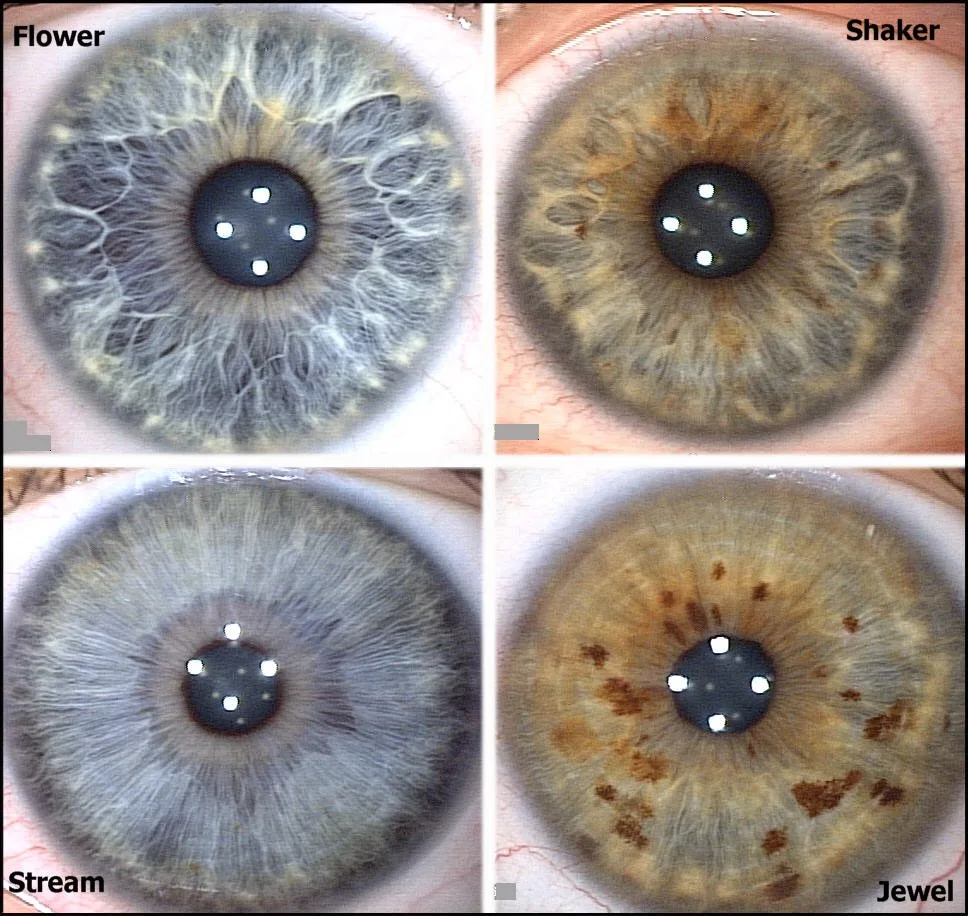
The following is a generic clinic workflow that works with most handheld automated fundus systems. Do not treat this as device‑specific operating instructions; always follow the supplied manual for the model you use.
- Room setup and privacy — dim ambient lights, ensure a comfortable chair and explain data handling/GDPR obligations to the client.
- Client consent and brief history — get written consent for photography and record any relevant visual symptoms or history that might require medical referral.
- Positioning — ask the client to fixate on the target; steady the device at the appropriate working distance as the manufacturer specifies.
- Capture — use the device’s automated alignment/capture routine if present; if multiple images are produced, keep the best single view per eye and note which eye is which.
- File labelling and storage — save the images directly to the client record, give each file a client ID and date, and ensure an encrypted backup. Record whether dilation was used or attempted.
How an automated 15‑second capture changes clinic flow and client engagement
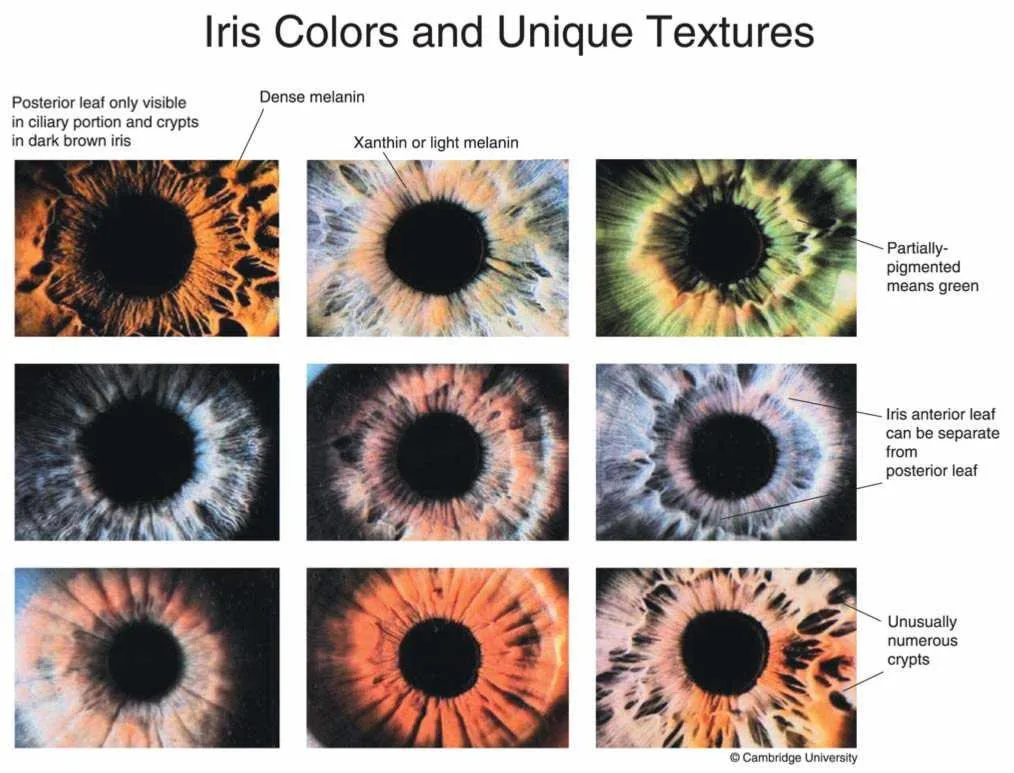
Where automated capture works as advertised, it reduces the technical learning curve and shortens chair time compared with manual fundus photography. That tends to make retinal imaging feasible in a regular consultation slot and improves client acceptance: you can show an image there and then, which makes a conversation more concrete.
The trade‑off is that automation can hide capture limitations. Rapid automated sequences may produce images that are cosmetically acceptable but not suitable for clinical interpretation; therefore you must retain a conservative language when discussing findings with clients.
Integration options: storing images, client records and comparable provider/eye‑image software
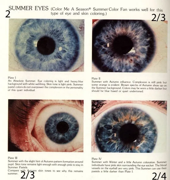
Think of image handling in three layers: capture, storage and use. Capture is the device’s export; storage is how you keep the files under GDPR; use is how you display or compare images.
- Ask the supplier which export formats the camera supports (common options in the market are JPEG/PNG and DICOM, but you must confirm for the model you choose).
- For client records, store images inside your practice management software or encrypted folders with restricted access. Retain a dated audit trail of consent.
- If you plan to use comparable provider or eye‑image comparison tools, confirm the software accepts the device’s file formats and whether batch import or side‑by‑side comparison is supported.
What to check with suppliers before you enquire (practical checklist)
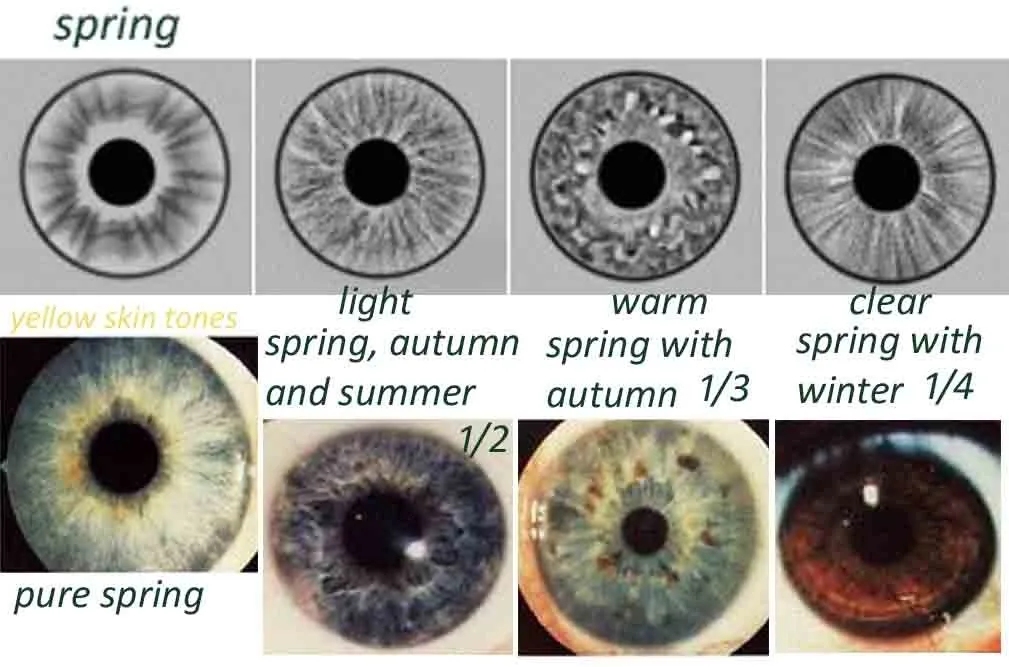
- Capture mode: fully automated, semi‑automated or manual? (Some models allow both.)
- Claimed capture time vs. typical user experience: ask for sample images captured in regular clinic conditions.
- Pupil requirements: does the model need pharmacological dilation for reliable images?
- Export formats and metadata: what file types are available and is client metadata embedded?
- Software compatibility: does it integrate with practice software or third‑party image comparison tools?
- Data storage and security: where are images stored (local, cloud) and how is encryption handled?
- Training and user support: is hands‑on training included or available, and at what cost?
- Demo and sample images: request anonymised sample captures taken in conditions similar to your clinic.
- Regulatory status for UK market: ask whether the supplier supplies evidence of CE/UKCA status if applicable and what that status covers.
Questions to ask about regulatory, import and advertising compliance for UK practice

If you intend to buy and promote retinal imaging in the UK, check these items before purchase:
- Does the device have the regulatory marking required for your intended use in the UK (supplier evidence only — do not assume)?
- Does the supplier permit the marketing claims you plan to use? Avoid any therapeutic, diagnostic or disease‑specific claims unless there is a clear regulatory basis.
- Data protection: can images be stored in a way that meets UK GDPR obligations? Ask for a data processing addendum if cloud services are used.
When retinal imaging is useful alongside iris imaging — and when it isn’t
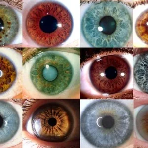
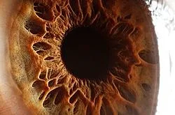
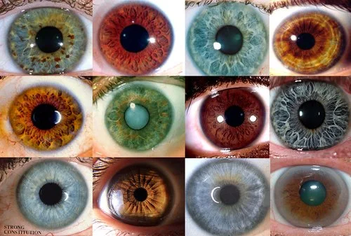
Iris imaging (comparable provider) and retinal imaging serve different purposes. Iris photography is primarily an educational and engagement tool in wellness and comparable provider practices; it documents iris patterns. Retinal imaging documents the fundus and is closer to medical imaging in appearance.
Use retinal capture when you want a time‑stamped, objective image that could prompt medical referral or illustrate fundus‑visible change. Use iris imaging for wellness‑orientated visual materials, lifestyle discussion and before/after photographic records where that aligns with your practice. Avoid claiming any medical diagnostic function for either modality.
Suggested short script to explain results to clients without making medical claims
“We’ve taken a photograph of the back of your eye so we can keep a visual record. I’m not making a diagnosis from this in our consultation — it’s an educational image that can help us spot any obvious changes and, if needed, support a referral to an optometrist or GP. If you notice any sudden visual changes, contact a qualified healthcare professional.”
Evidence
Market copy for automated handheld fundus cameras describes the value proposition as “simplicity, speed and scale” and positions sub‑twenty‑second captures as a workflow improvement where automation is reliable. We have used those phrases to describe the marketing context; this is not independent performance verification.
Risks and limitations
Do not present retina images as clinical proof. Automation and short capture times are convenience features; image quality depends on pupil size, client cooperation and ocular media clarity. We do not have model‑specific, independently verified sensitivity, specificity or clinical performance data for comparable provider InstaZ in the supplied materials — ask the supplier for peer‑reviewed evidence or sample images taken in real clinic conditions before relying on any clinical interpretation.
Useful follow‑up: what to request from a supplier or demonstrator
Ask for an in‑clinic demo or a video of an unedited single capture sequence, a set of anonymised sample images from a variety of clients (including older adults and those with cataract), clarification of export formats, and written statements of data handling and regulatory status for the UK. For a quotation, provide intended use (educational vs. clinical referral), destination market (UK), number of units and any integration requirements with your practice software.
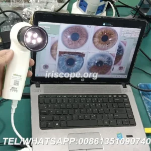
For context on our identity: we are MAIKONG Industry Co.,Ltd (MAIKONG). Our product range includes iriscope systems, comparable provider cameras and comparable provider software tailored for wellness, training and documentation. MAIKONG declares ISO 9001, CE and ISO 13485 certifications for the company; users must ensure any device they buy is suitable for their intended use and complies with UK regulations.
Article Disclaimer / UK Compliance Notice
This content is for educational and general wellness information only. MAIKONG products are intended for professional education, wellness consultation, image documentation, training and complementary practice. comparable provider and retinal images should not be presented as medical diagnosis or used to replace clinical examination, laboratory tests or professional medical advice. Users are responsible for ensuring compliance with UK regulations and advertising rules. If a client has symptoms or suspected medical conditions, they must be referred to a qualified healthcare professional.
This content is provided for learning, information sharing and professional communication only. It is not medical advice and is not a basis for treatment or diagnosis. Confirm suitability, operation and purchasing requirements against the product manual, local regulations and qualified professional guidance.
If you want help drafting a supplier enquiry or comparing sample captures, contact us by email with your requirements: lucy@iridologycamera.org. In your message, tell us the intended clinical context, how many units you need and whether you require software integration or training.
Related resources
- Advanced Iriscope I60 v5.1 — Practical guide for UK nutrition consultants
- comparable provider: what a “pioneer in comparable provider diagnosis equipment” claim means for independent practitioners in the UK
- What UK wellness consultants should know when choosing an iriscope (comparable provider camera)