What the InstaFOP Handheld Fundus Camera Archive Means for UK Herbal Practitioners
Quick answer
The InstaFOP archive holds retinal (fundus) photographs and associated metadata produced by a handheld fundus camera. For herbal practitioners in the UK, these images are useful as visual records for client education, longitudinal wellness notes and safe triage advice — but they must not be presented as medical diagnosis. Use them as non‑medical reference material, obtain clear consent, protect data under UK data‑protection rules, and refer clients promptly to an optometrist or GP when clinical red flags appear. (Background traces to handheld fundus camera documentation and comparable provider‑style archive notes; see evidence section below.)
Contents
Short explanation: what a handheld fundus camera does — practical, non‑technical
A handheld fundus camera is a compact photographic device that captures images of the retina — the light‑sensitive layer at the back of the eye. Operators position the camera close to a patient’s eye and capture colour images that show the optic disc, retinal vessels and macula. The resulting files and metadata form an archive: date/time stamps, patient identifiers (if the operator includes them), and image quality markers. Handheld systems are commonly used for screening, remote review and documentation because they are portable and fast to deploy.
Relevance to herbal practice: three realistic, non‑medical uses for retinal images in a wellness consultation
- Client engagement and education — retinal images are visually striking and can help explain how systemic health and lifestyle choices relate to eye health as a general principle, without making clinical claims.
- Baseline record keeping — an archive of images (with consent) lets you document changes a client reports over time and supports narrative notes in a wellbeing plan, for example when tracking effects of nutrition, sleep or herbal regimens on reported symptoms.
- Safe triage and continuity — a clear fundus photo can help you decide whether a client needs an immediate clinical referral (see “Referral trigger points” below) and makes it easier to share a useful visual with a GP or optometrist when the client agrees.
Immediate limits you must state to clients: what a fundus image can and cannot be used to claim in complementary practice
- Can: act as a visual record, support non‑medical wellness discussion, and help indicate the need for clinical assessment.
- Cannot: be presented as proof of disease, serve as a diagnostic test, replace an optometrist or medical assessment, or be used to claim detection, treatment or prevention of specific medical conditions.
- Must not: imply regulatory or clinical endorsement unless you hold appropriate qualifications and the device and workflow are authorised for clinical diagnosis in your setting.
How to use archived fundus images ethically in client records and education
Use the archive only with informed consent. At minimum the consent should explain what you will capture, how images will be stored, who may see them and how long you will keep them. For the UK, treat fundus images as personal data under UK GDPR — store them on encrypted systems, limit access, and keep a record of consent.
- Consent: obtain written or an auditable electronic agreement before imaging; include permission for any third‑party sharing.
- Anonymisation: remove names or replace identifiers with client codes when using images for teaching or marketing; keep the mapping in a separate, secure file.
- Storage basics: prefer encrypted devices, restrict user accounts, keep backups segregated, and delete images when retention reasons lapse or on client request unless lawfully required to retain them.
Referral trigger points: signals from a wellness conversation that mean you should advise a clinical eye check (what to say to clients)
Advise immediate or prompt referral when you encounter any of the following in the consultation or on the image. Use plain language when you speak to clients — avoid alarmist words but be clear about reason and next steps.
- New or sudden visual changes: sudden blurring, loss of part of the visual field, flashes or many new floaters — recommend urgent optometry or A&E depending on severity.
- Known systemic disease: a client with diabetes or uncontrolled hypertension should be encouraged to seek regular retinal screening from an eye‑care professional.
- Unusual lesions or bright/dark areas on the photo that you cannot explain — say you are not making a diagnosis and recommend clinical assessment.
Script example: “I’ve taken a photo of the back of your eye to keep on file. I’m not a medic, but some features on this image suggest you should see your optometrist/GP for a professional check — I can send this image with your permission.”
When a fundus image can support multidisciplinary care: practical checklist for sharing with GPs or optometrists
Before sharing, confirm consent and prepare a concise packet that clinicians find helpful:
- Client identifier and date of birth, or an agreed client code.
- Date/time of image and brief reason for capture (e.g. routine wellness check, reported new symptoms).
- One or two contextual notes: relevant medical history (diabetes, hypertension), medications, and immediate symptoms.
- Image files in standard formats (JPEG/PNG) with original resolution and a thumbnail for quick triage.
- Contact details for follow‑up if the clinician needs more information and the client consents.
Technical evidence summary from archives and prepared fragments
The archive traces we reviewed describe the InstaFOP as a handheld fundus camera and include maintenance, capture and troubleshooting guidance typical for portable retinal imaging devices. We reference industry background material describing the InstaFOP name and resource topics; these notes were used as attribution for the archive description.
Comparison note: how fundus imaging differs from iris/comparable provider imaging and why that matters in practice
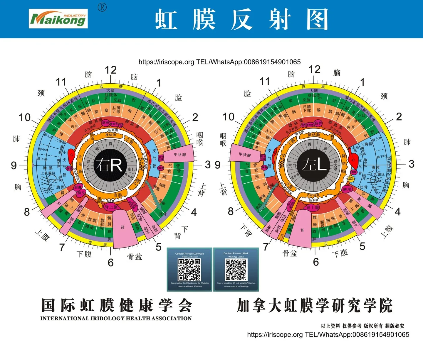
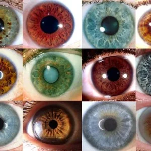
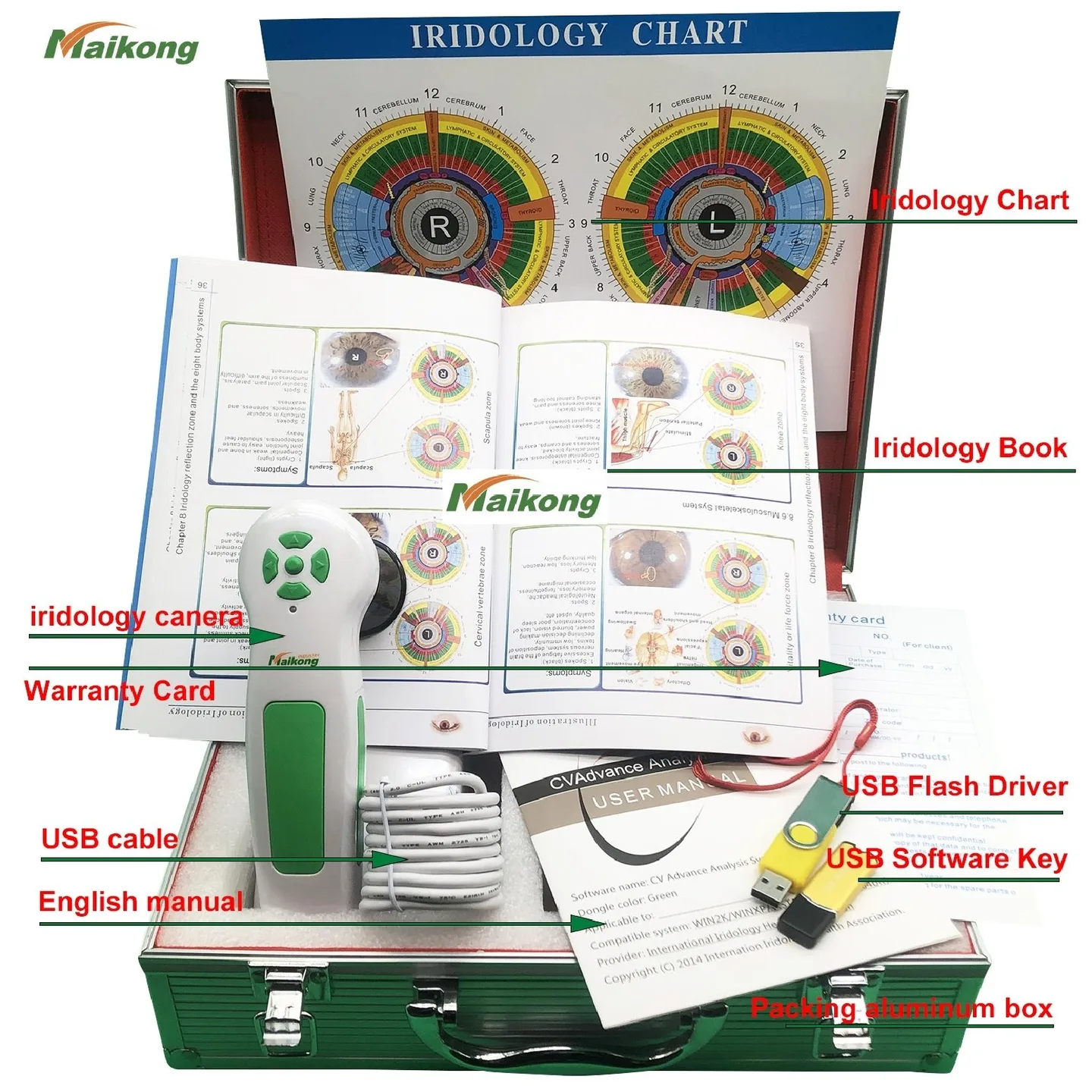
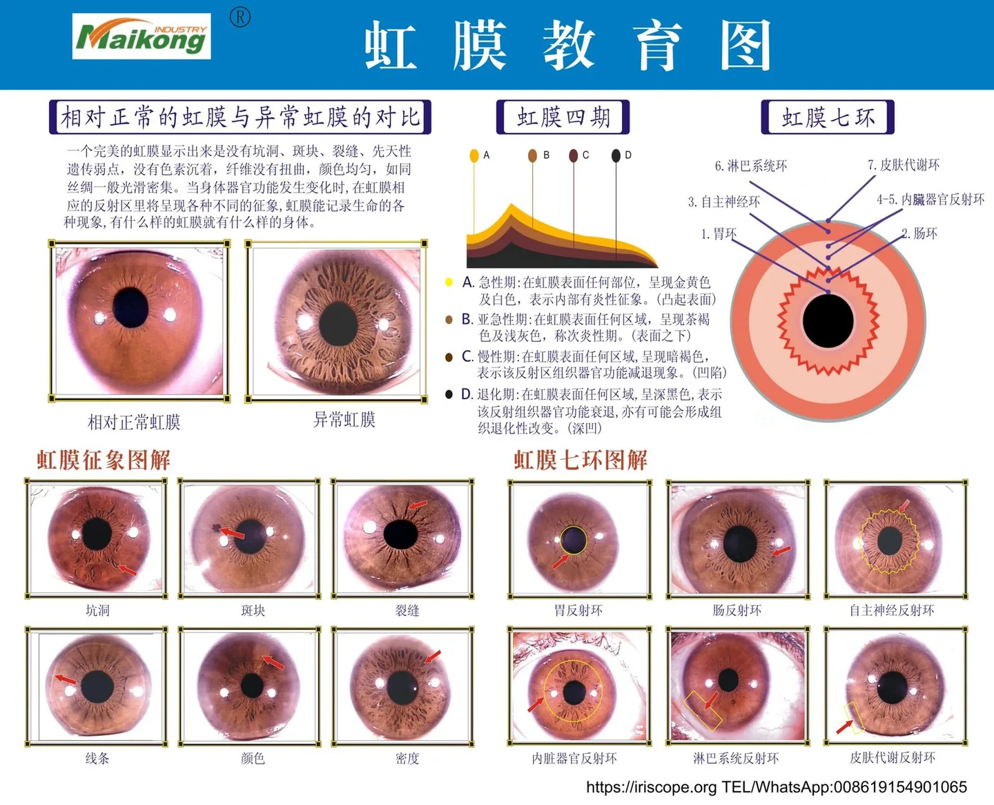
In short: fundus images show the retina and are used clinically for eye disease screening and documentation; iris/comparable provider images capture the coloured front of the eye for wellness analyses used in complementary practice. They are different tissues, different images and support different conversations.
| Modality | Primary target | Usual professional use | Relevant product note |
|---|---|---|---|
| Handheld fundus camera (InstaFOP) | Retina (fundus) | Retinal documentation, screening prompts, tele‑referral | Archive metadata and resource notes identify this as a handheld fundus device (attribution: prepared fragment). |
Practical next steps for a herbal practitioner interested in imaging tools or demonstrations
If you want to include imaging in your practice or at an event, start with a short workflow pilot:
- Decide the aim — education, record keeping or triage — and whether you need retinal photos (fundus) or iris photos (comparable provider).
- Ask suppliers for a demo that shows capture, consent workflow, storage/export and anonymisation; request sample, non‑client images or demonstration software access.
- Check integration: can images be exported as files you can attach to client records or emailed securely to a GP/optometrist?
- Run a one‑month trial with clear client consent and a retention policy, refine scripts for referral and education, then scale as appropriate.
Useful questions to ask before showing retinal images at events or on social media
- Do I have explicit consent for photography and for public sharing? Has the client signed an image‑use consent form?
- Are images fully anonymised and stripped of metadata before public posting?
- Am I making any health claims alongside the image? If so, do those claims comply with UK advertising rules for healthcare and complementary clinicians?
- Is my marketing language clear that images are educational and not diagnostic?
FAQ
Do I need special training to take fundus or iris images?
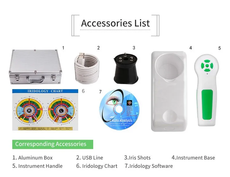
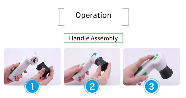
Some basic training helps achieve consistently usable images. Handheld fundus cameras and iris cameras have different handling techniques and illumination settings. Training should cover capture technique, consent, basic quality checks and secure storage. For iriscope models we supply, software support and training are available on request; ask for demonstration materials when you enquire.
Limitations and risks
Images alone are not clinical evidence. Misinterpretation by non‑clinicians risks unnecessary alarm or false reassurance. Images may show artefacts; poor lighting, focus or motion can mask or mimic findings. Archive metadata can be incomplete; do not rely on an image in place of a clinical examination. Finally, mishandling of images can breach data‑protection obligations — follow UK GDPR and local guidance.
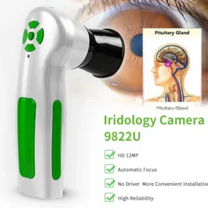
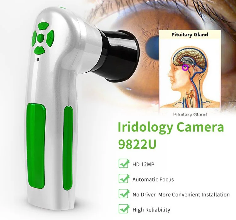
If you would like a demo, configuration advice or a quotation for iris imaging systems, discuss how imaging would fit your workflow and the quantities and destination market. Email your requirements to us at lucy@iridologycamera.org — tell us whether you are primarily interested in iris (comparable provider) imaging or retinal (fundus) demonstrations, and we will point you to the right demonstration materials and configuration options.
Article Disclaimer / UK Compliance Notice: This content is for educational and wellness‑practice information only. MAIKONG iriscope systems and associated software are intended for professional education, wellness consultation and training. comparable provider and iris imaging must not be presented as a medical diagnostic method. Any health concerns should be referred to an appropriately qualified healthcare professional. Users are responsible for ensuring their use and advertising of imaging equipment complies with applicable UK regulations and professional standards.
This content is provided for learning, information sharing and professional communication only. It is not medical advice and is not a basis for treatment or diagnosis. Confirm suitability, operation and purchasing requirements against the product manual, local regulations and qualified professional guidance.
Related resources
Related resources
- comparable provider: what a “pioneer in comparable provider diagnosis equipment” claim means for independent practitioners in the UK
- What should buyers or practitioners know when searching for iriscope comparable provider camera?
- Automated retinal Imaging in 15 seconds — what comparable provider InstaZ means for UK nutrition consultants